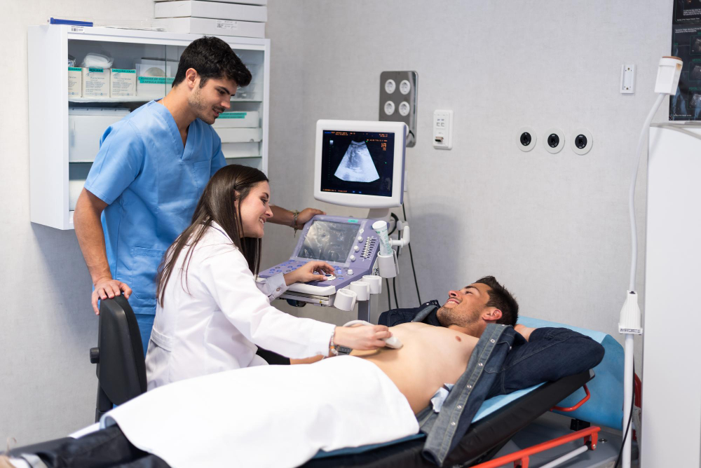
2D Echo

At Relief Hospital, we offer 2D Echo (two-dimensional echocardiography), an advanced, non-invasive imaging technique that plays a vital role in diagnosing and managing various heart conditions. By utilizing high-frequency sound waves, the 2D Echo produces real-time images of the heart, allowing healthcare professionals to assess its structure and functionality effectively.
Key Benefits of 2D Echo
Comprehensive Heart Assessment: The 2D Echo provides critical information about the heart’s chambers, valves, and blood flow patterns, helping to identify any abnormalities or structural defects.
Diagnosis of Cardiac Conditions: This imaging modality is essential in diagnosing various conditions, including heart valve diseases, cardiomyopathies, and congenital heart defects. It is also useful for detecting fluid around the heart (pericardial effusion) and measuring heart size and function.
Monitoring Treatment Progress: For patients with existing heart conditions, the 2D Echo is instrumental in monitoring changes over time, enabling healthcare providers to adjust treatment plans based on the heart’s evolving status.
The Procedure
The 2D Echo procedure involves:
- Preparation: Patients are typically asked to lie down and may need to expose their chest for the ultrasound.
- Image Capture: A transducer is placed on the chest, emitting sound waves that create images of the heart in real-time. The process is painless and usually takes about 30 to 60 minutes.
- Interpretation: Our experienced cardiologists analyze the generated images to provide a comprehensive report on the heart’s health and recommend further management if necessary.
Commitment to Quality Care
At Relief Hospital, we are committed to providing exceptional cardiovascular care. Our state-of-the-art equipment and skilled team ensure accurate diagnostics, allowing patients to receive the most effective treatments for their heart health.
